 中央研究院 生物化學研究所
中央研究院 生物化學研究所
中央研究院 生物化學研究所
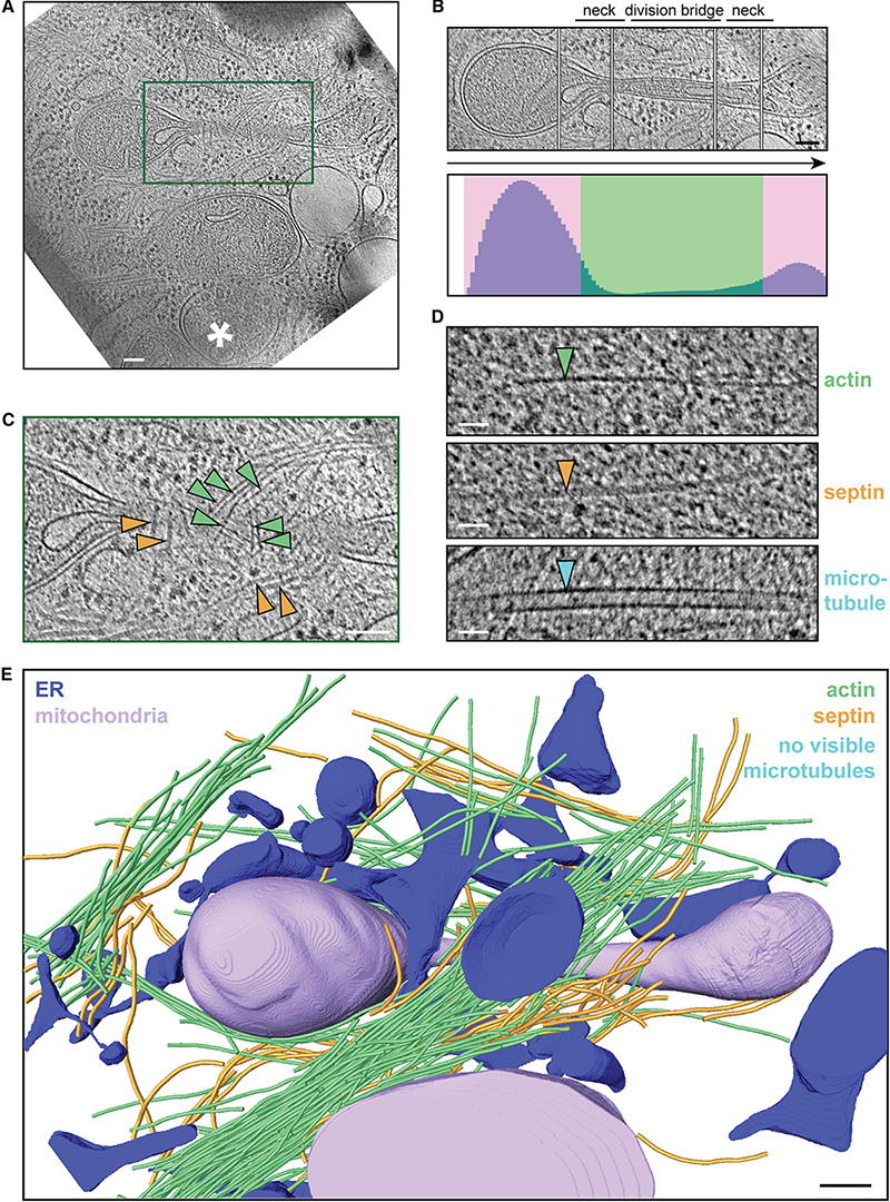
Mitochondria adapt to changing cellular environments, stress stimuli, and metabolic demands through dramatic morphological remodeling of their shape, and thus function. Such mitochondrial dynamics is often dependent on cytoskeletal filament interactions. However, the precise organization of these filamentous assemblies remains speculative. Here, we apply cryogenic electron tomography to directly image the nanoscale architecture of the cytoskeletal-membrane interactions involved in mitochondrial dynamics in response to damage. We induced mitochondrial damage via membrane depolarization, a cellular stress associated with mitochondrial fragmentation and mitophagy. We find that, in response to acute membrane depolarization, mammalian mitochondria predominantly organize into tubular morphology that abundantly displays constrictions. We observe long bundles of both unbranched actin and septin filaments enriched at these constrictions. We also observed septin-microtubule interactions at these sites and elsewhere suggesting that these two filaments guide each other in the cytosolic space. Together, our results provide empirical parameters for the architecture of mitochondrial constriction factors to validate/refine existing models and inform the development of new ones.