 中央研究院 生物化學研究所
中央研究院 生物化學研究所
中央研究院 生物化學研究所
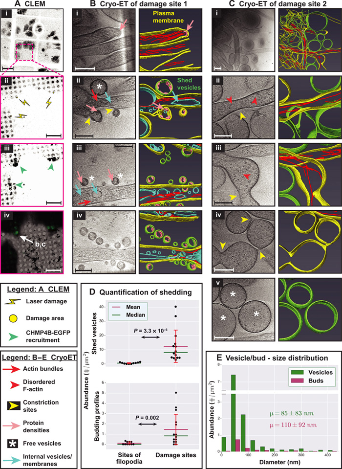
Cryo-electron tomography (cryo-ET) provides structural context to molecular mechanisms underlying biological processes. Although straightforward to implement for studying stable macromolecular complexes, using it to locate short-lived structures and events can be impractical. A combination of live-cell microscopy, correlative light and electron microscopy, and cryo-ET will alleviate this issue. We developed a workflow combining the three to study the ubiquitous and dynamic process of shedding in response to plasma membrane damage in HeLa cells. We found filopodia-like protrusions enriched at damage sites and acting as scaffolds for shedding, which involves F-actin dynamics, myosin-1a, and vacuolar protein sorting 4B (a component of the 'endosomal sorting complex required for transport' machinery). Overall, shedding is more complex than current models of vesiculation from flat membranes. Its similarities to constitutive shedding in enterocytes argue for a conserved mechanism. Our workflow can also be adapted to study other damage response pathways and dynamic cellular events.