 中央研究院 生物化學研究所
中央研究院 生物化學研究所
中央研究院 生物化學研究所
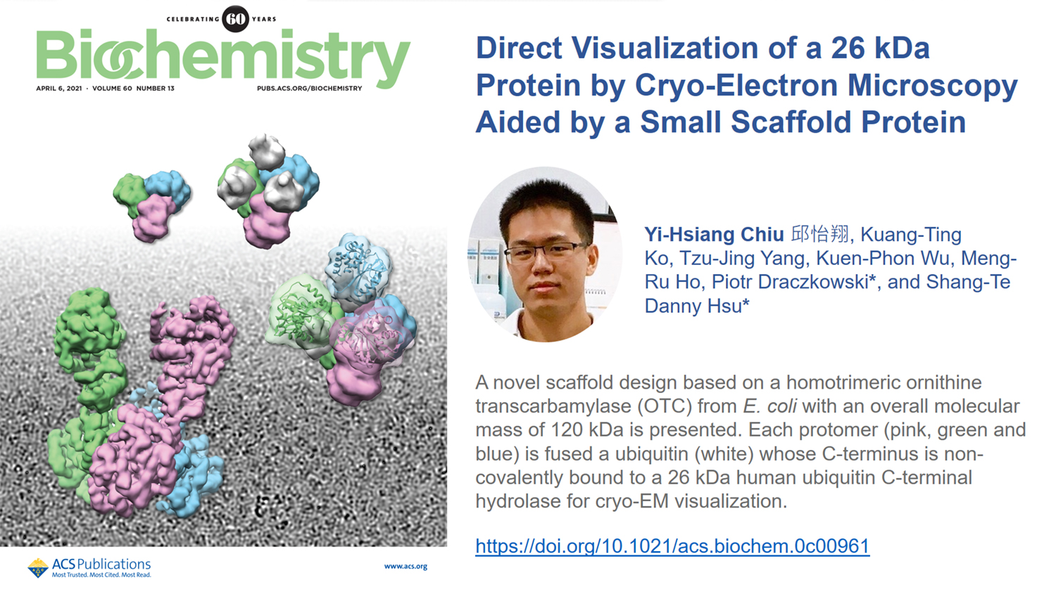
Cryo-electron microscopy (cryo-EM)-based structure determination of small proteins is hindered by the technical challenges associated with low signal-to-noise ratios of their particle images in intrinsically noisy micrographs. One solution is to attach the target protein to a large protein scaffold to increase its apparent size and, therefore, image contrast. Here we report a novel scaffold design based on a trimeric helical protein, E. coli ornithine transcarbamylase (OTC), fused to human ubiquitin. As a proof of principle, we demonstrated the ability to resolve a cryo-EM map of a 26 kDa human ubiquitin C-terminal hydrolase (UCHL1) attached to the C-terminus of ubiquitin as part of the trimeric assembly. The results revealed conformational changes in UCHL1 upon binding to ubiquitin, namely, a significant displacement of α-helix 2, which was also observed by X-ray crystallography. Our findings demonstrated the potential of the trimeric OTC scaffold design for studying a large number of ubiquitin interacting proteins by cryo-EM.