 中央研究院 生物化學研究所
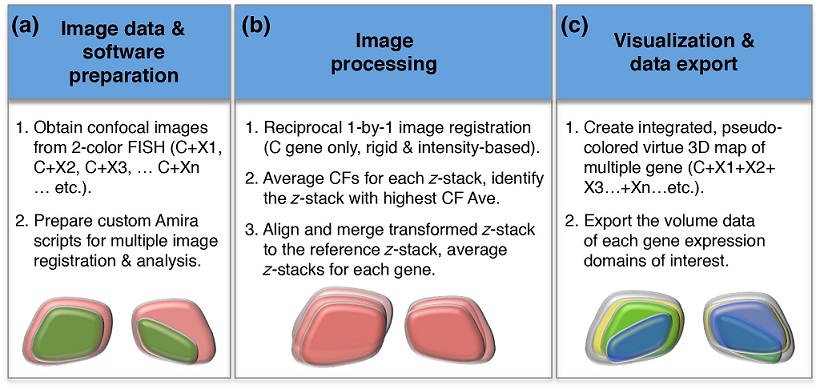
The conserved bilateral habenular nuclei (HA) in vertebrate diencephalon develop into compartmentalized structures containing neurons derived from different cell lineages. Despite extensive studies demonstrated that zebrafish larval HA display distinct left-right (L-R) asymmetry in gene expression and connectivity, the spatial gene expression domains were mainly obtained from two-dimensional (2D) snapshots of colorimetric RNA in situ hybridization staining which could not properly reflect different HA neuronal lineages constructed in three-dimension (3D). Combing the tyramide-based fluorescent mRNA in situ hybridization, confocal microscopy and customized imaging processing procedures, we have created spatial distribution maps of four genes for 4-day-old zebrafish and in sibling fish whose L-R asymmetry was spontaneously reversed. 3D volumetric analyses showed that ratios of cpd2, lov, ron, and nrp1a expression in L-R reversed HA were reversed according to the parapineal positions. However, the quantitative changes of gene expression in reversed larval brains do not mirror the gene expression level in the obverse larval brains. There were a total 87.78% increase in lov+ nrp1a+ and a total 12.45% decrease in lov+ ron+ double-positive neurons when the L-R asymmetry of HA was reversed. Thus, our volumetric analyses of the 3D maps indicate that changes of HA neuronal cell fates are associated with the reversal of HA laterality. These changes likely account for the behavior changes associated with HA laterality alterations.
