 中央研究院 生物化學研究所
中央研究院 生物化學研究所
中央研究院 生物化學研究所
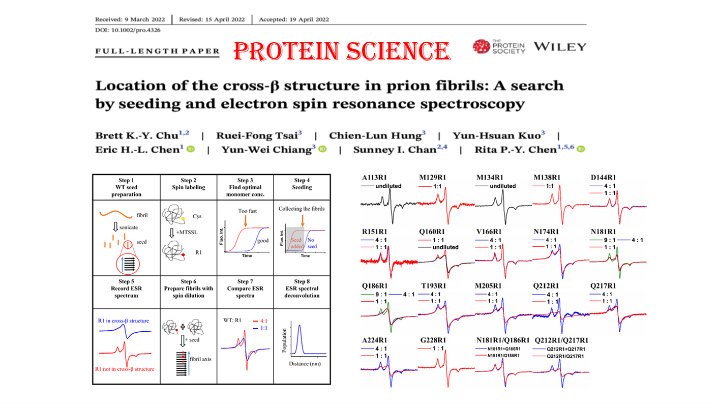
Prion diseases are transmissible fatal neurodegenerative disorders spreading between humans and other mammals. The pathogenic agent, prion, is a protease-resistant, β-sheet-rich protein aggregate, converted from a membrane protein called PrPC . PrPSc is the misfolded form of PrPC and undergoes self-propagation to form the infectious amyloids. Since the key hallmark of prion disease is amyloid formation, identifying and studying which segments are involved in the amyloid core can provide molecular details about prion diseases. It has been known that the prion protein could also form non-infectious fibrils in the presence of denaturants. In this study, we employed a combination of site-directed nitroxide spin-labeling, fibril seeding, and electron spin resonance (ESR) spectroscopy to identify the structure of the in vitro-prepared full-length mouse prion fibrils. It is shown that in the in vitro amyloidogenesis, the formation of the amyloid core is linked to an α-to-β structural transformation involving the segment 160-224, which contains strand 2, helix 2, and helix 3. This method is particularly suitable for examining the hetero-seeded amyloid fibril structure, as the unlabeled seeds are invisible by ESR spectroscopy. It can be applied to study the structures of different strains of infectious prions or other amyloid fibrils in the future.