Facility at NTU campus
LOCATION
IBS R108, 206, 208, 310, 400
CONTACT
Sheng-Wei Lin sanway@gate.sinica.edu.tw +886-2-23665540
Description

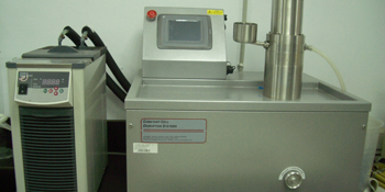
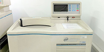
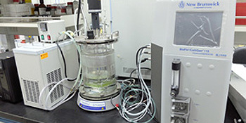
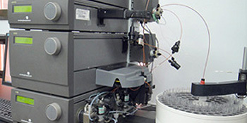
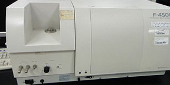
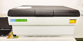
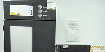
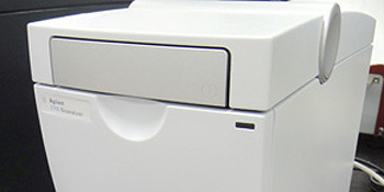
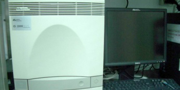
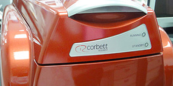
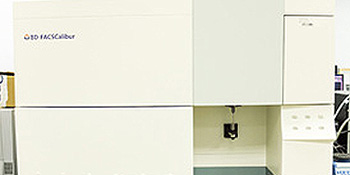
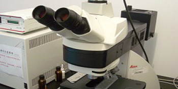
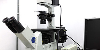
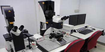
The IBS Core Facility is located at the Institute of Biochemical Sciences (IBS), College of Life Science, National Taiwan University. It is jointly established by the Institute of Biological Chemistry (IBC), Academia Sinica and IBS. The facility provides technical support for investigators of IBC and IBS in instrument operation training, scheduling, workshops, user certification, trouble shooting and instrument maintenance. The major instruments of the core facility include: Flow Cytometry (BD FACSCalibur), Confocal Microscope System (Leica SP5), Real-Time PCR Systems (Bio-Rad CFX Connect and Qiagen Rotor-Gene), Upright and Inverted Microscope Systems (Leica DM6000B and OLYMPUS), Fluorescence/Luminescent CCD Image Analyzer (Vilber Lourmat Fusion FX 7 and ThermoFisher iBright FL1500), Bioanalyzer System (Agilent 2100), Protein Purification System (GE ÄKTA™), Fluorescence Spectrophotometer (Hitach F-4500), Radioisotope Counter (PerkinElmerTri-Carb 2900TR), Cell Disruption System (Constant Systems Ltd TS 0.75Kw), Cryostat (Leica CM 1950) and Ultracentrifuge (Beckman XL-100K).

In addition, the IBS Core Facility also houses a Biomolecular Interaction Analyzer (GE BIAcore T200) provided by the Technology Commons (Techcomm) of the NTU College of Life Science, which are open to IBC/IBS users.

1. Constant Systems Ltd TS 0.75Kw Cell Disruption System 
Location: IBS R108

Our cell disruption system is capable of processing all types of samples (algae, bacteria, fungi, mammalian cells and yeasts). Disruption pressures ranging from 1K psi to 40K psi can be set and displayed digitally. It is capable of continuously processing large quantities of cultured cells. The disruption head is surrounded by a unique cooling jacket to prevent overheating even after several cycles of cell disruption.

2. Beckman XL-100K Ultracentrifuge 
Location: IBS R108

This ultracentrifuge has a maximum rotational speed of 100,000 rpm and is equipped with different rotors (VTI 65, SW 28, 41Ti, 55Ti, Type 45Ti, 55.2Ti and 70Ti).

3. New Brunswick BioFlo/CelliGen 115 Benchtop Fermentor 
Location: IBS R400

It is an integrated control station with a color touch-screen interface, built-in pumps, gas flow controllers, pH/DO, and foam/level controllers. Using this fermentor, large quantities of bacteria (3 litres) can be grown to very high concentrations. This fermentor automatically detects and controls all of the above parameters for the optimization of bacterial growth.

4. GE ÄKTA™ Protein Purification System 
Location: IBS R400

ÄKTA™ Protein Purification System is an instrument for fast protein liquid chromatography (FPLC). Our system is equipped with four pumps for running four different solutions simultaneously, a full wavelength UV detector, a pH detector, a conductivity detector and an automated fraction collector.

5. Hitach F-4500 Fluorescence Spectrophotometer 
Location: IBS R400

The intense 150 watt xenon source of the F-4500 fluorescence spectrophotometer provides maximum light energy over the entire range of 200-900 nm and allows acquisition of spectra in less than one second. Wavelength scan speeds of up to 30,000 nm/min provide rapid scans for both excitation and emission. Its pre-scan function ensures automatic and fast selection of the optimum excitation and emission wavelengths for your unknown samples.

6. PerkinElmer Tri-Carb 2900TR Radioisotope Counter 
Location: IBS R206-1

Liquid scintillation analyzer Tri-Carb 2900TR (Perkin Elmer) is a computer-controlled benchtop liquid scintillation analyzer for detecting small amounts of α, β and γ radioactivity. Its TR-LSC (Time-Resolved Liquid Scintillation Counting) feature ensures high sensitivity and the low background counting of liquid scintillation samples.

7. Vilber Lourmat Fusion FX 7 Fluorescence/Luminescent CCD Image Analyzer) 
Location: IBS R400

Fusion FX7 is a CCD image acquisition system can be used to detect trans-illumination/epi-illumination white light, UV and fluorescent lights (red, green and blue). It is dedicated to the capture of chemiluminescence and fluorescence digital pictures from gels or membranes and digitally quantifies the intensity of your samples.

8. Agilent 2100 Bioanalyzer System 
Location: IBS R400

The Agilent 2100 Bioanalyzer is a microfluidics-based platform for sizing, quantification and quality control of DNA, RNA, proteins and cells. Results are delivered within 30-40 minutes in automated, high quality digital data. The advantages offered by this system include improved data precision and reproducibility, short analysis times, minimal sample consumption and improved automation.

9. Applied Biosystems 7300 Real-Time PCR 
Location: IBS R400

Real-time PCR is a laboratory technique based on the polymerase chain reaction, which is used to amplify and simultaneously quantify a targeted DNA molecule. In our core facility, there are two real-time PCR systems. The Applied Biosystems 7300 Real-time PCR features a tungsten halogen lamp that illuminates all sample wells in the 7300 system. Fluorescence emission can be passed through four filters and is collected by a CCD camera. The Qiagen Rotor-Gene Real-time PCR features an LED lamp and a single optical detection pathway. In this system, all samples are kept at the same temperature by spinning the samples in a chamber of moving air.

10. Qiagen Rotor-Gene Real-Time PCR 
Location: IBS R400

Real-time PCR is a laboratory technique based on the polymerase chain reaction, which is used to amplify and simultaneously quantify a targeted DNA molecule. In our core facility, there are two real time PCR systems. The Applied Biosystems 7300 Real-time PCR features a tungsten halogen lamp that illuminates all sample wells in the 7300 system. Fluorescence emission can be passed through four filters and is collected by a CCD camera. The Qiagen Rotor-Gene Real-time PCR features an LED lamp and a single optical detection pathway. In this system, all samples are kept at the same temperature by spinning the samples in a chamber of moving air.

11. BD FACSCalibur Flow Cytometry 
Location: IBS R400

Flow cytometry is a microscope and counter-based technology employed in cell counting, sorting and biomarker detection by suspending cells in a stream of fluid and passing them through an electronic detection apparatus. It allows simultaneous multiparametric analysis of both the physical and chemical characteristics of cells. Our instrument is equipped with two lasers for the excitation at 488 and 633nm. Combined with four fluorescence channels, both forward and side scatter data can be collected. It can be used for immunophenotyping, absolute cell counting, stem cell analysis and DNA analysis, etc.

12. Leica DM6000B Upright Microscope System 
Location: IBS R411

These microscopes are used to capture imaging for cell research. It features a motorized z-focus and a motorized and coded 7x nosepiece. The motorized stage and focus drive offers the convenience of automatic adjustment, allowing the automated positioning of Z levels. These microscopes allow several kinds of detection modes, including bright field, phase contrast, differential interference contrast and fluorescence imaging.

13. OLYMPUS IX71 Inverted Microscope System 
Location: IBS N401

These microscopes are used to capture imaging for cell research. It features a motorized z-focus and a motorized and coded 7x nosepiece. The motorized stage and focus drive offers the convenience of automatic adjustment, allowing the automated positioning of Z levels. These microscopes allow several kinds of detection modes, including bright field, phase contrast, differential interference contrast and fluorescence imaging.

14. Leica SP5 Confocal Microscope System 
Location: IBS N401

Confocal microscopy is an optical imaging technique used to increase optical resolution and contrast of a micrograph by using a spatial pinhole to eliminate out-of-focus light in specimens. This Confocal Microscope is equipped with four laser lines, namely 405nm diode, Argon laser (458, 488, 514nm), 543nm laser and 633 nm laser. It provides many applications including cell imaging study, colocalization, FRAP, FRET, time scan, Lamda scan and Z-axis scan, etc. In our facility, the confocal system is equipped with an upright microscope and an inverted microscope.