Institute of Biological Chemistry, Academia Sinica
Research Highlights
Put a lid on it: the structure that allows the NlpI-Prc complex to degrade MepS in Escherichia coli
By Cindy Lee
A new structural study, published online on November 15 in Nature Communications, demonstrates how a lipoprotein adaptor and a protease form a three-sided docking cradle to mediate protein degradation in the E. coli outer membrane. The research team led by Dr. Chung-I Chang, Associate Research Fellow of the Institute of Biological Chemistry, has revealed the coordination between a homodimer of the lipoprotein adaptor NlpI and two molecules of the periplasmic PDZ-protease Prc by using X-ray crystallography, a technique in which molecules in the form of crystals are shot with X-rays to determine the molecules’ three-dimensional structures. NlpI and Prc form a complex in the outer membrane to degrade the outer membrane lipoprotein MepS, a process necessary for regulating the bacterial growth and stationary phases.
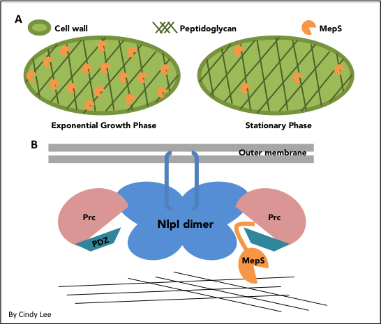
Inside the cell wall, a mesh-like structure called the peptidoglycan (PG) provides structural support to the cell (Figure A). In order for the PG to expand and accommodate bacterial growth, hydrolases like MepS are necessary to cleave the cross-links and make room for new PG material.
The NlpI-Prc complex is important for regulating MepS activity (Figure B). Chang’s group determined the structure of the complex to be a butterfly-shaped homodimer of NlpI with a circular Prc molecule on each side of the homodimer. NlpI has tetratricopeptide repeats (TPRs) on the outer edges of the dimer that are important for interacting with Prc. The C-terminal domain (CHD) and N-terminal domain (NHD) of Prc form a bowl-shaped body that is attached with a hinge to a lid-like PDZ domain, which provides a complementary catalytic pocket.
Although Prc is responsible for cleaving MepS, it alone is not enough to bind MepS. It turns out that another NlpI TPR on the edge of the homodimer is responsible for the interaction with MepS, recruiting MepS to the valley between NlpI and Prc. There, the Prc hinge residues allow PDZ to detect and degrade MepS.
In determining the structure of the NlpI-Prc complex, Chang’s group has highlighted the differences between Prc and other proteases. The unique bowl-and-lid structure of Prc promotes cleavage of MepS and downstream affects on PG activity. In light of the importance of PG activity in bacterial cell growth and stability, these findings illuminate the role of lipoproteins in regulating cell wall enzymes and suggest new targets for antibacterial agents.

 Institute of Biological Chemistry, Academia Sinica