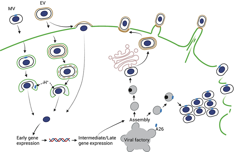
Poxviruses are double-stranded DNA viruses that represent the largest known highly pathogenic viruses infecting humans. They undergo dramatic morphological changes during their maturation process, resulting in structural differences between each virion, and their surface is decorated with more than a dozen randomly distributed surface proteins that facilitate viral entry. These are the main reasons poxviruses have eluded high-resolution structure determination. Over the last three decades, cryo-EM has developed into a mature technology that can increasingly overcome such problems of structural heterogeneity through advances in microscope technology and image processing algorithms. Here, we discuss the essential current modalities in cryo-EM, which promise to solve the structure of poxviruses in parts and as entire virions at near-atomic resolution. With a focus on cryo modalities, we provide an overview of methods, including volume microscopy by plasma ion beam milling, focused ion beam lamella preparation, subtomogram averaging, and single particle averaging. Protocols for poxvirus propagation, purification, and imaging by cryo-EM are presented. This chapter is aimed at experts and nonexpert researchers to help facilitate entry into the structural biology of this critical field in virology.

 Institute of Biological Chemistry, Academia Sinica
