Cell Biology
Histopathology
LOCATION
Rm416
CONTACT
Hsiao-Fang Nien mifiy111@gate.sinica.edu.tw +886-2-27855696,4161
Description

The IBC Histopathology Core Facility was established in early 2013. The mission is to serve the need of IBC researchers in histology and pathology analyses in order to promote the studies of animal models and human diseases. The Core lab currently houses several essential equipments including an automatic tissue processor, a tissue embedding centre, a rotary microtome for routine paraffin sections, and a cryostat for frozen sections.

Requests for histopathology services should be sent to Hsiao-Fang Nien via E-mail.

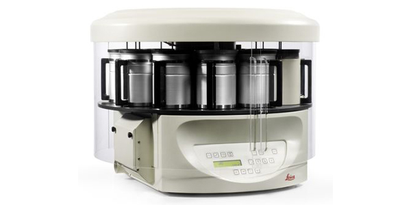
1. Semi-enclosed benchtop tissue processor, Leica TP1020

Leica TP1020 automatic tissue processor has 12 reagent containers, and it could store 9 programs. Through the processes of dehydration, wash, and rinse, the fixative tissue samples could maintain appropriate quality for embedding.

 
2. Modular tissue embedding center, Leica EG1150

Leica 1150 tissue embedding center could dispense and fill the melting paraffin in the molds which contain tissues/specimens. Besides, it could move the products upon the cold plate for cooling, and could gain paraffin embedding blocks easily.

 
3. Manual rotary microtome for routine sectioning, Leica RM2235

Leica RM2235 rotary microtome could orient and fix the specimen in horizontal and vertical direction which provides proper paraffin sections for microscopic observation.

 
4. Cryostat/Microtome (Leica CM1950 )

The Leica CM1950 cryostat provides a stable low temperature environment and maintains the quality of cryosections for microscopy.