Cell Biology
Imaging
LOCATION
Rm416
CONTACT
Jung-Kun Wen lunkung@gate.sinica.edu.tw +886-2-27855696,4160
Description

The Bio-Imaging Core Facility is located at Room 416, Institute of Biological Chemistry, Academia Sinica. 
For instrument education and training information please refer to https://sites.google.com/view/bicf

This facility provides the following services for IBC researchers: 
Design of bio-imaging-related experimental methods. 
Assist in the selection of suitable imaging systems, image processing, and image analysis. 
Provide biological imaging-related application knowledge and course training. 
One-stop bio-imaging experiment and analysis services.

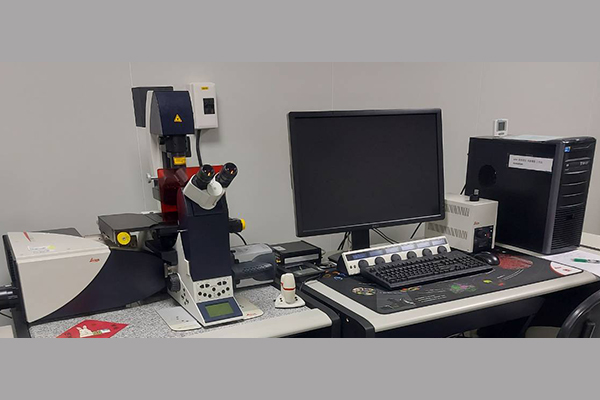
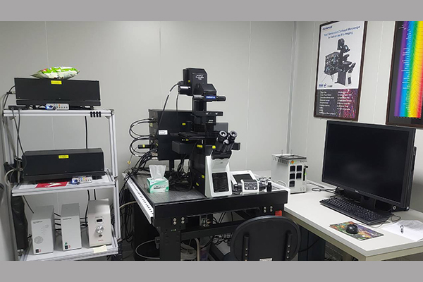
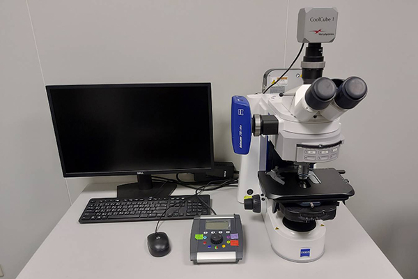
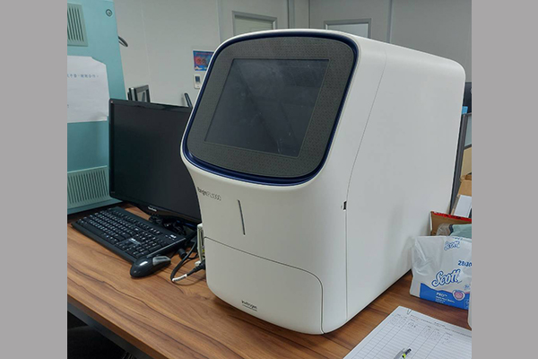
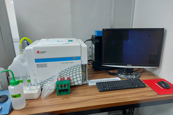
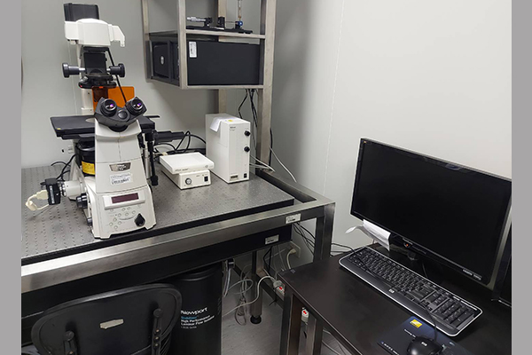
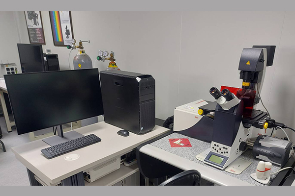
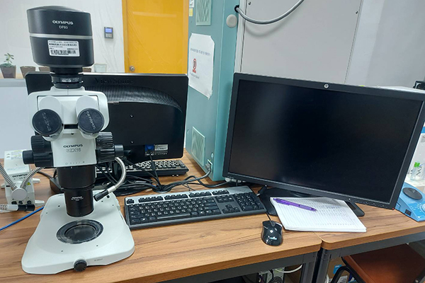
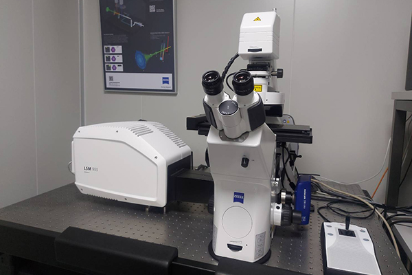
Imaging systems provided at this facility include: 
Laser-scanning confocal microscopes 
Fluorescence microscope 
High-content screening station 
Flow cytometers 
Bioluminescence and Chemiluminescence Imager.

Policies & Pricing

Acknowledgement of the Core Facility 
All users of the Imaging Core Facility should acknowledge the facility in publications and presentations that include data generated in the facility. Please also acknowledge the person who services imaging analysis services. We also appreciate receiving an electronic version of publications that include data gathered in our facility.

Acknowledgment template 
We thank the Bio-Imaging Core Facility at the Institute of Biological Chemistry, Academia Sinica, for the technical support in image acquisition and analysis.

1. Leica SP5 X Inverted Confocal Microscope

Equipped  
Laser: 405, 470-670 white laser  
Motorized XY Scanning Stage  
Z-axis micro-vibration stage 

Application  
Live Cell Imaging  
Functional imaging experiments  
FRAP (Fluorescence recovery after photobleaching)  
FRET (Fluorescence Resonance Energy Transfer)  
Fluorescence Colocalization

2. Olympus FV3000RS Inverted Confocal Microscope

Equipped 
Laser : diode laser 405, 445, 488, 514, 561, 594 and 640 
Motorized XY Scanning Stage 
High Sensitivity-Spectral Detector 
Resonant scanner 
Silicone Objectives 

Application 
Live Cell Imaging 
Functional imaging experiments 
FRAP (Fluorescence recovery after photobleaching) 
FRET (Fluorescence Resonance Energy Transfer) 
Fluorescence Colocalization

3. Olympus scan^R high-content screen station

Equipped 
Olympus IX83 Automated inverted microscope 

Application 
Cell counting 
Intracellular transport 
Translocation 
Cell proliferation 
Bacterial and viral infection assays 
Cell-cycle analysis 
Protein localization and colocalization 
Live cell assays 
Fluorescence analysis in tissue sections 
Cell migration

4. Zeiss Axio Imager.A2 Microscope

Equipped 
ZEISS Axiocam 208 color camera 
CoolCube 1Digital High Res. CCD Camera 

Application 
Image acquisition and reporting 
Fluorescence analysis in tissue sections 
Karyotyping

5. Fujifilm Luminescent Image Analyzer LAS4000

Equipped 
Trans-illumination white light and UV 
Epi-illumination white light, red, green and blue fluorescence. 

Application 
Chemiluminescence imaging 
Fluorescence imaging

6. iBright FL1000 Imaging Systems

Equipped 
9.1 megapixel cooled 16-bit CCD camera 
Invitrogen Smart Exposure technology 
Advanced automated features 
Green LED-based transilluminator 

Application 
Protein gel and membranes 
Nucleic acid gel 
Chemiluminescence imaging 
Fluorescence imaging

7. Beckman CytoFLEX

Equipped 
Laser : 405,488,638nm 
8 channels for fluorescence detection 

Application 
Immunophenotyping 
Absolute counting 
Residual white blood cell enumeration 
Stem cell analysis 
DNA analysis

8. Nikon Ti-E Microscope

Application 
Image acquisition and reporting 
Fluorescence analysis in tissue sections

9. SRRF Stream+ Super-Resolution Microscopy System

‘SRRF-Stream+’ is a real-time super-resolution microscopy functionality that super-charges the imaging performance of your standard microscope.

Application 
Image acquisition and reporting 
Fluorescence analysis in tissue sections

10. Research stereomicroscope with a dual-CCD system, Olympus SZX16 with DP80

Olympus SZX16 with DP80 acquire bright-field and fluorescence images of the same field. Meanwhile, the slim LED transmitted light illuminator provides brightfield, contrast-enhancing oblique and darkfield illumination, it performance superior quality image for research purpose.

11. ZEISS LSM 900 with Airyscan 2 Inverted Confocal Microscope

Equipped  
Laser : diode laser 405, 488, 561, and 640 
Motorized XY Scanning Stage  
GaAsp ultra-high sensitivity sensor  
32 channel matrix GaAsp sensor  
10x Plan-Apochromat objective  
20x Plan-Apochromat objective  
40x LD C-Apochromat Water objective  
63x Plan-Apochromat Oil objective  
DIC observation kit  

Application  
Super Resolution (AiryScan)  
Colocalization  
3D imaging  
Time-lapse imaging,Photo Bleaching (FRAP, FLIP, FRET, Photoactivation, Photoconversion)  
Linear unmixing